An atrioventricular block, in general, occurs when there is a conduction delay between the atria and ventricles of the heart. A second-degree atrioventricular (AV) block type I is also known as Wenckebach or Mobitz type I. This rhythm occurs at the AV node, where conduction of an electrical impulse is impaired. Impulses from the AV node progressively slow until an impulse fails to conduct to the ventricles (the impulse is then considered “blocked”); this cycle typically repeats following the non-conducted beat. A second-degree AV block type I rhythm is often bradycardic; in an adult patient, bradycardia is widely classified as a heart rate less than 60 beats per minute, though a heart rate less than 50 beats per minute is the standard for some clinicians when managing patient care for symptomatic bradycardia.
Image credit: https://www.vdh.virginia.gov/content/uploads/sites/23/2016/05/CAR-508.pdf
A patient with a second-degree AV block type I may have the following signs or symptoms:
This heart block is generally considered benign. Most commonly, there is little hemodynamic disruption, and patients are largely asymptomatic. This heart block is less likely to transition to a third-degree heart block (a complete heart block), which is considered a rhythm at high risk for the development of ventricular tachycardia, asystole, and sudden cardiac death.
Potential causes of this rhythm include:
It is essential to identify the difference between a second-degree AV block type I (Wenckebach or Mobitz type I) and type II (Mobitz type II) as the management approach to each is different. Patients with a second-degree AV block type I may be asymptomatic. If the patient with bradycardia has a second-degree AV block type I (or other bradyarrhythmia) and is symptomatic, it is crucial to determine if presenting signs or symptoms are due to the slowed heart rate; this would then be considered unstable bradycardia.
Signs and symptoms of unstable bradycardia may include:
If the patient is not showing any signs or symptoms of poor perfusion (as described above), then the patient can be observed and monitored for any potential change in their clinical condition. However, if it is determined that an adult patient with bradycardia is symptomatic, unstable (showing signs or symptoms of poor perfusion), and has a pulse, then the Advanced Cardiovascular Life Support (ACLS) Adult Bradycardia Algorithm should be initiated by the healthcare provider to guide evaluation and treatment.
According to the ACLS Adult Bradycardia Algorithm, an initial step a healthcare provider should take in caring for a patient is completing an ACLS Primary Assessment to identify and treat underlying potential causes for the patient’s condition. This includes managing the patient’s airway, providing supplemental oxygen if needed, determining the patient’s cardiac rhythm, and monitoring their vital signs. Additionally, intravenous access should be established, a 12-lead ECG should be obtained if possible, and the healthcare provider should assess and consider possible reversible causes (Hs & Ts) for the patient’s clinical condition. The patient’s clinical condition and their response to treatment may necessitate the healthcare provider to utilize multiple interventions concurrently while continuing to monitor for any changes to prevent the patient’s condition from deteriorating and developing cardiac arrest.
If there are no immediately reversible causes and the patient is demonstrating signs or symptoms of poor perfusion as a result of a bradyarrhythmia, the ACLS Adult Bradycardia Algorithm indicates that atropine may be administered. Atropine is considered a first-line treatment; a dose of 0.5 mg intravenously and can be repeated every 3-5 minutes until a maximum dose of 0.4mg/kg (3mg in a 75kg patient) has been administered. Symptomatic patients with a second-degree AV block type I generally respond to atropine; permanent cardiac pacing for patients with a second-degree AV block type I is uncommon.
If the patient with a bradyarrhythmia such as a second-degree AV block type I continues to demonstrate signs and symptoms of unstable bradycardia after the use of atropine, transcutaneous pacing (TCP) may be utilized. Transcutaneous pacing transmits an electrical stimulus from an external power source (such as a defibrillator with a pacing function) through electrodes applied strategically to the surface of the patient’s skin. ACLS providers are able to perform TCP. If the patient is conscious, they should be sedated prior to initiating this intervention if possible, as this intervention can be painful. Transcutaneous pacing should be utilized as an intervention to bridge treatment until the patient can be transferred to a higher level of care for expert consultation.
A second-degree AV block type I bradyarrhythmia is often benign; many patients are asymptomatic. If this rhythm was induced by a medication, the rhythm disturbance often reverses after the medication’s cessation or a dosing modification - this would warrant management through expert consultation. Symptomatic patients with a second-degree AV block type I generally respond to the use of atropine. Patients with this bradyarrhythmia who are symptomatic should be transferred to a higher level of care for expert consultation, continued monitoring, and management.
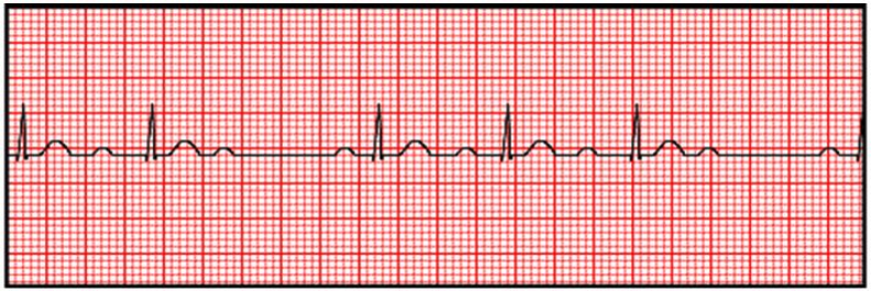
Understanding the difference between second-degree AV blocks type I and type II is key to providing accurate and effective patient care. A second-degree AV block type I occurs at the AV node of the heart. On an ECG, the PR interval gradually lengthens until the QRS complex ‘drops’ (more P waves are present than QRS complexes). A repeating pattern of one less QRS complex than P waves in a series is typical (where a P:QRS ratio occurs within a series of 3:2 or 4:1, as an example).
A second-degree AV block type II (Mobitz Type II) occurs when electrical impulses are intermittently blocked below the AV node. An ECG for this rhythm would reflect that P waves are regular, the PR interval is consistent, and (similar to a second-degree AV block type I) there will be more P waves than QRS complexes though the dropped QRS complex occurs unexpectedly in a second-degree AV block type II.
The key to distinguishing these two rhythms is to evaluate the PR interval and determine whether the PR interval is progressively prolonging (type I) or constant (type II). A consideration that reinforces the importance of being able to distinguish between these two rhythms is that there is a decreased likelihood of a second-degree AV block type I transitioning to a worsening AV block (a complete heart block) as compared to a second-degree AV block type II, which has an increased potential for deteriorating to a life-threatening infranodal level AV block or asystole.
This is covered in depth in the Pediatric Advanced Life Support (PALS) course.
Achieving and maintaining these certifications ensures that you are a knowledgeable and skillful healthcare provider ready to respond and care for patients experiencing life-threatening cardiac emergencies. To earn this certification, you’ll need to master common ECG rhythms and the appropriate procedures to respond to each.
The American Medical Resource Institute (AMRI) will prepare you for completing your Basic Life Support (BLS), ACLS, or PALS certification exam(s) using a multidisciplinary approach to teaching that incorporates challenging and innovative learning opportunities with the flexibility you need to complete your certification coursework and exam at any time, from any device, in one sitting or over time.
Would you like to work through scenario-based case studies to test your knowledge? Click here for ACLS and PALS case studies based on pre-hospital, hospital, or outpatient settings.
Did you know that by completing your certification course through AMRI that you gain access to an expanded library of case studies and practice tests that are only available to registered students? Register for a BLS, ACLS, or PALS certification course here.
When you register for your exam, you get access to exclusive practice tests and case studies.