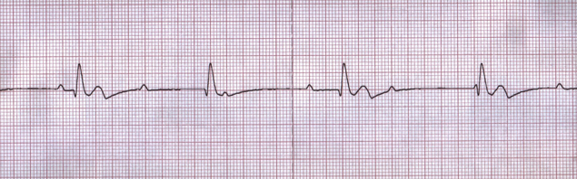
Pulseless electrical activity (PEA) is defined as the presence of cardiac electrical activity with organized or partially organized cardiac rhythms without a palpable pulse. Pulseless electrical activity is formerly known as electromechanical dissociation (EMD). During PEA, the heart is unable to move adequate blood volume to maintain systemic perfusion.
Rhythms such as ventricular fibrillation and pulseless ventricular tachycardia are not associated with pulseless electrical activity (despite being pulseless) as they tend to respond to defibrillation. Asystole is considered a separate rhythm than pulseless electrical activity.
Signs or symptoms of pulseless electrical activity may include:
Pulseless electrical activity occurs due to an event that takes place within the respiratory, cardiovascular, or gastrointestinal systems of the body which contributes to insufficient force generated by the heart following electrical depolarization. This insufficient force results in a weakened ability of the heart to contract, which can lead to a worsening cascade of pathophysiological changes such as hypoxia, acidosis, and decreased vagal tone. Further impairment of the contractility of the heart results, despite the presence of electrical activity within the heart, and mechanical activity of the heart becomes insufficient. The risk for clinical mortality is increased in patients experiencing pulseless electrical activity.
Causes of pulseless electrical activity can be differentiated into primary and secondary causes. Primary pulseless electrical activity is associated with cardiac causes (such as cardiac arrest), whereas secondary pulseless electrical activity is due to noncardiac causes. Secondary pulseless electrical activity is due to potential reversible causes, also known as the Hs & Ts:
Hypoxia and hypovolemia are two of the most common and potentially reversible causes of pulseless electrical activity. Medications may increase susceptibility for the development of pulseless electrical activity. For example, patients who use beta-blockers or calcium channel blockers are at an increased risk for pulseless electrical activity as a result of decreased contractility of the heart due to altered intracellular calcium levels.
In caring for an adult patient, the healthcare provider should initially complete the Basic Life Support (BLS) Assessment, the Advanced Cardiovascular Life Support (ACLS) Primary Assessment (such as managing the patient’s airway, providing supplemental oxygen if needed, evaluating their cardiac rhythm, monitoring vital signs, establishing intravenous (IV) or intraosseous (IO) access, evaluating H’s and T’s, etc.), and the ACLS Secondary Assessment (a focused medical history). For adult patients who have no pulse and an organized cardiac rhythm (indicative of pulseless electrical activity), the ACLS Cardiac Arrest Algorithm is utilized to guide treatment following the asystole/PEA pathway. Pulseless electrical activity is considered a non-shockable rhythm.
Interventions for managing pulseless electrical activity are focused on tending to the underlying cause of cardiac arrest rather than converting the cardiac rhythm. Patient outcomes are dependent on identifying and correcting an underlying cause of this rhythm and performing effective cardiopulmonary resuscitation (CPR). The ability to recognize when a heart rate is tachycardic or bradycardic, regular or irregular, as well as when a QRS complex is wide or narrow may be helpful in providing care for the patient with pulseless electrical activity.
If a return of spontaneous circulation (ROSC) occurs and a pulse and an organized cardiac rhythm are present, transition to post-cardiac arrest care for the patient. Extending resuscitative efforts may be warranted if the patient has experienced potentially reversible causes of cardiac arrest, such as a drug overdose or hypothermia. However, it may be necessary to evaluate whether resuscitation efforts should be continued contingent on a combination of factors, such as the clinical condition of the patient, the duration of time that CPR has been performed, the patient’s response to resuscitative efforts, and the setting in which the patient is being cared for (in-hospital versus out-of-hospital) in addition to respective protocols and policies. The duration of time of resuscitative efforts alone is not an appropriate measure to determine if efforts should cease. Follow the protocols and policies in place by your institution when evaluating whether to terminate resuscitative efforts.
Pulseless electrical activity can be separated into pseudo-PEA or true-PEA. Pseudo-PEA occurs when the patient has no palpable pulse, yet identifiable pressures within the aorta and weak ventricular contractions are present. True-PEA occurs when electrical impulses are firing without cardiac contractions.
Diagnostic equipment such as ultrasound can be a useful adjunct tool to discern whether a patient is experiencing true-PEA or pseudo-PEA. However, ultrasound may not be available, depending on the setting in which the healthcare provider is practicing. Without ultrasound, this determination is otherwise subjective and complicated by the potential difficulty in assessment due to the experience level of the responding healthcare provider, unknown circumstances surrounding the cardiac arrest (such as an unwitnessed event), and/or anatomical considerations of the patient (such as obesity).
Did you know that if you register for your certification course through AMRI that you will have access to an expanded library of case studies and practice tests that are otherwise unavailable to the public?
American Medical Resource Institute (AMRI) prepares you for successful completion of your BLS, ACLS, or pediatric advanced life support (PALS) certification(s) so that you may effectively respond to a life-threatening emergency and apply your knowledge and skills to improve potential patient outcomes. AMRI provides rigorous, innovative, and multidisciplinary opportunities to maximize your learning and offers a flexible and convenient learning environment. You can complete your coursework and exam according to your schedule, on any device.
Would you like to review five things you need to know about PEA? Review them here.
Are you searching for scenario-based case studies to reinforce and test your knowledge? Click here for ACLS and PALS case studies that are tailored to individual practice settings.
Ready to register for your BLS, ACLS, or PALS certification or renewal? Register here.
When you register for your exam, you get access to exclusive practice tests and case studies.